Retina and vitreous

Retina doesn't fit in a free-text field.

A detachment is drawn, not described. The full retina, already built.

Create my account

14-day trial. No card needed to sign up.

View clinical demos
A smiling female physician shows a tablet to an older patient in an ophthalmology practice. Illustrative image generated with AI.

A retina consultation

Chief complaint

Guided mode
FloatersFlashesBlurred vision
Demo PatientFirst visit · retina consultation

You pick the symptoms and the note builds itself.

Fundus map

12 hours, 3 zones
TearHemorrhageDrusenNeovessels
Right eyeTear at 2 o'clock, mid-periphery

The fundus is drawn, not described.

Macular OCT

Trend
Central thickness412 µm
Pre-check358 µm
ClassificationDME, standardized

The study is compared with the previous one.

Plan

Intravitreal injection
Plan with intravitreal injectionRight eye · intravitreal

Supply deducted from inventory

The supply is deducted automatically.

Review and sign

You sign
Retina noteDemo Patient · ready for review

Sign note

Note signed

AI proposes. You review and sign.

Illustrative example, fictitious data.

How it's captured

The fundus is drawn. The note writes itself.

12-hour, 3-zone retina chart, right eye Diagram of the fundus of the right eye divided into posterior pole, equator, and periphery, with the 12 clock hours marked. Three findings are shown: a horseshoe tear at 11 o’clock in the periphery, prior photocoagulation around the tear, and lattice degeneration between 1 and 2 o’clock. 36912 Superior Inferior Nasal Temporal Posterior pole Equator Periphery
  • Horseshoe tear · 11 · periphery
  • Prior photocoagulation · 10 to 12
  • Palisading · 1 to 2 · periphery
What the system writes into the note on its own OD: horseshoe tear at 11 o'clock in the periphery, with prior photocoagulation from 10 to 12. Lattice degeneration from 1 to 2 o'clock in the periphery. Posterior pole unremarkable.
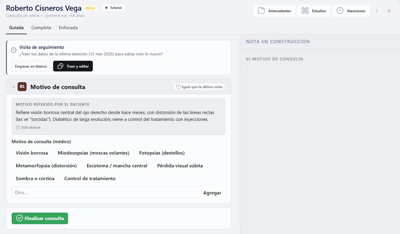
Illustrative screenshot of the iGalenus clinical demo: first-visit retina consultation with a demo patient. It shows guided mode with the chief complaint, symptom options for the physician and the note being built.

Illustrative screenshot of the clinical demo. The patient is a demo patient.

Your progress, charted

Trend in central macular thickness and visual acuity across visits Illustrative sample chart: central macular thickness drops from 520 to 330 microns while visual acuity rises from 0.3 to 0.6. 600500400300µm0.80.60.40.2decimalV1V2V3V4V5Successive visits Central macular thicknessVisual acuity
  • Central macular thickness · 520 → 330 µm
  • Visual acuity · 0.3 → 0.6 decimal
Illustrative example

This is the trend the system charts on its own, from what you already capture at each visit. The values are an illustrative example, not a real patient.

Illustrative sample chart with made-up data. Does not correspond to any patient.

From dictation to signature

AI proposes. You review and sign.

From dictation to signed note Four steps in order: dictation, structured note, note review before signing, and the doctor's signature. Artificial intelligence proposes; the doctor confirms and signs. DictationYou speakwhile you examineStructured noteThe AI proposeseach fieldNOM-004 reviewBefore signing,score from 0 to 100Doctor's signatureYou confirmand sign From dictation to signed note Four steps in order: dictation, structured note, note review before signing, and the doctor's signature. Artificial intelligence proposes; the doctor confirms and signs. DictationYou speakwhile you examineStructured noteThe AI proposeseach fieldNOM-004 reviewBefore signing,score from 0 to 100Doctor's signatureYou confirmand sign
AI proposes; you confirm and sign. Once signed, the note is immutable.

Try the Retina and vitreous note with your own data.

What costs you time today

The fundus map is drawn on a loose sheet that never makes it back into the record.

The OCT lives on the machine; comparing it with the one from three months ago is done from memory.

The intravitreal injection is given, but the schedule and re-injection are noted separately.

At iGalenus

What the Retina and vitreous record includes.

12-hour, 3-zone retina map

Tears, hemorrhages, drusen, and neovessels with standard symbols; written up automatically in the note.

Macula and OCT, charted

Macular thickness and findings per eye, on a curve over time.

Standardized classification

NPDR, proliferative retinopathy, DME and AMD: you choose, you don't type.

Plan with intravitreal injection

Drug, eye and schedule on a plan line, with re-injection follow-up.

Supplies are deducted automatically

What you apply in the note comes out of inventory at that same moment.

Retina plan in one click

Anti-VEGF, laser, panretinal photocoagulation, vitrectomy and surveillance.

Studies being compared

Wipe, side by side, flicker, and 2×2 grid, with synchronized zoom.

AI-assisted interpretation

AI drafts the OCT report; you review and sign it.

And everything else is still there.

Scheduling and rescheduling by WhatsApp AI dictation Prescriptions with QR Payments, invoicing, and inventory NOM-004 before signing Optical Patient portal

Questions about Retina and vitreous

What we're usually asked.

Is the retina map drawn with a finger on a tablet?

Yes. The map works with a finger, digital pen or mouse. Stamps and sectors are placed on a 12-hour, 3-zone diagram, and the system translates the drawing into text within the clinical note.

Can I put the intravitreal injection in the patient's plan?

Yes. In the plan you write the intravitreal injection with drug, eye and schedule, and pick the follow-up (for example, 4 weeks for the re-injection). It stays in the signed note.

Does it connect with my OCT?

Studies are uploaded directly from the technician workstation connected to the device. The system recognizes the file type, suggests the eye and date, and you confirm.

Your Retina and vitreous record, ready to go.

Create my account

14-day trial. No card needed to sign up.

See pricing
Create my account